Shockwave Therapy in the Racehorse: What the Equine Literature is Teaching Us
And why it matters for human musculoskeletal practice too
There’s a quiet revolution happening in the equine veterinary literature, and most of us in human musculoskeletal medicine aren’t reading it. We probably should be.
In December 2025, Frontiers in Veterinary Science published a systematic review by Qiu and colleagues at Northeast Agricultural University in Harbin, China, drawing together more than two decades of research on extracorporeal shockwave therapy in horses. It is the most comprehensive synthesis I’ve seen of the equine ESWT evidence base, and reading it carefully reveals something striking: the horses are teaching us things about shockwave that we cannot easily learn from human studies alone.
For 38 years I have been practising osteopathic medicine, and for the last eleven years I have been delivering focused extracorporeal shockwave therapy as a core part of my clinical work. In that time, the conversations with colleagues — and increasingly with patients who have done their own reading — keep returning to the same two questions. Does it really matter whether the device is focused or radial? And is shockwave actually doing anything beyond providing temporary analgesia?
The equine literature gives the clearest answers I have seen to both.
Why horses are unusually informative
The horse is an extraordinarily useful model for human musculoskeletal medicine, and not for sentimental reasons. Equine athletes carry musculoskeletal loads comparable to elite human athletes — Thoroughbreds gallop at 40 mph carrying their own weight plus a rider, generating forces that would destroy the tendons and ligaments of any less specialised mammal. The tendon and ligament structures are scaled similarly to ours. The injury patterns map across species with remarkable fidelity.
Proximal suspensory desmitis in the horse behaves like high hamstring tendinopathy and plantar fasciitis in our patients. Superficial digital flexor tendinitis behaves like Achilles tendinopathy. Bone spavin behaves like advanced ankle and subtalar osteoarthritis. The navicular bone, sitting behind the hoof capsule, is anatomically and biomechanically analogous to a deep, attenuated target in human practice — the deep gluteal tendons, the high hamstring origin, the deep posterior compartments of the leg.
Critically, the equine literature can do experimental work we simply cannot do in humans for ethical reasons. Collagenase-induced tendon lesions, serial histology at predefined intervals, biopsy of treated tissue, controlled comparison of high-energy and low-energy protocols against placebo — these are routine in horses and impossible in our species. The mechanistic detail in the equine literature is therefore far richer than what we have in our own.
When equine experimental data converges with human randomised controlled trial outcomes on the same therapeutic combinations, the translational signal is unusually robust. That convergence is exactly what we now have.
Finding one: focused outperforms radial in deep tissue
The head-to-head comparisons in the equine literature are striking, and they are consistent.
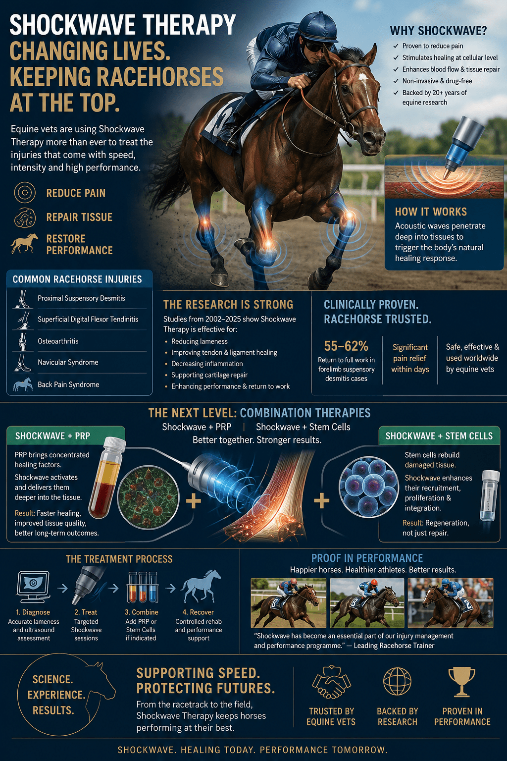
In chronic proximal suspensory desmitis — one of the most common causes of lameness in racehorses — Lischer and colleagues at the University of Zurich treated 56 horses with focused electrohydraulic shockwave (0.15 mJ/mm², 2,000 pulses, three sessions at three-week intervals). At six months, 61.8% of forelimb cases had returned to full work; at one year, 55.9% were still working fully. Crowe and colleagues at the Animal Health Trust in Newmarket used radial pressure wave therapy in the same indication: at six months, 53% of forelimb cases were sound and back to full work — and the gap widened in the more challenging hindlimb cases.
In navicular syndrome the comparison is even sharper. Blum and colleagues at the University of Giessen treated 42 horses with focused shockwave. The application site mattered enormously — 80% of horses treated through the heel bulb became sound, against only 47.4% treated through the sole (attributed to the higher keratin content of the sole attenuating shockwave energy on its way to the navicular bone). McClure and colleagues at Iowa State reported lameness improvement in 81% of non-blinded and 56% of blinded assessments with a single high-energy focused session. Brown and colleagues at Michigan State, using radial pressure wave therapy in the same indication, reported no improvement in lameness at all.
The mechanism behind this is straightforward physics. Focused shockwaves are generated by piezoelectric, electromagnetic, or electrohydraulic systems that concentrate acoustic energy to a defined focal point at depth, with minimal energy loss along the way. Radial pressure waves, generated ballistically, attenuate substantially during transmission and deliver their highest energy at the skin surface, falling off rapidly with depth.
For superficial work — myofascial trigger points, surface enthesopathy, soft tissue tone modulation — radial therapy has a role and serves it well. But for any structure sitting at depth — the navicular bone behind the hoof capsule, the proximal suspensory under multiple fascial layers, the thoracolumbar facets, the deep gluteal tendons in our patients, the high hamstring origin, the subchondral bone of an arthritic hip or ankle — focused is the evidence-supported choice. The two technologies are complementary, not interchangeable.
Finding two: shockwave is not just an analgesic device
The second finding to emerge consistently from the equine work is that shockwave’s clinical effect is structural as well as symptomatic. It is a mechanotransductive stimulus that recruits multiple parallel signalling pathways, not simply a pain-modulating device.
Bolt and colleagues at Louisiana State University showed that radial shockwave applied to equine palmar digital nerves caused extensive separation of myelin layers in medium-to-large myelinated axons (5–15 µm diameter), with no changes in small-diameter or non-myelinated axons. The injury is to myelin, not to axons or Schwann cell bodies, and crucially it is reversible. This is the cellular basis for shockwave’s selective analgesic effect — large-fibre pain transmission is dampened while small-fibre function is preserved.
But the analgesia is only one strand. Caminoto and colleagues at São Paulo State University used a collagenase-induced suspensory desmitis model — three sessions of focused ESWT significantly increased fine collagen fibril density, mitochondrial content, and extracellular matrix components at the lesion. McClure and colleagues showed the same in a forelimb model: ultrasound and histology returned to near-normal after three focused sessions. Kersh and colleagues demonstrated increased angiogenesis at the tendon injury site and raised extracellular matrix secretion by tenocytes after ESWT in collagenase-induced SDFT.
Frisbie and colleagues at Colorado State conducted a randomised controlled trial comparing focused ESWT, polysulfated glycosaminoglycan, and placebo in surgically-induced equine osteoarthritis. ESWT reduced lameness within two weeks and maintained improvement throughout the 70-day study — outperforming PSGAG. Kawcak and colleagues, in a companion paper, showed that ESWT raised serum osteocalcin and CTX-I (markers of bone turnover) and increased synovial proteoglycan. The bone-side mechanism of shockwave in osteoarthritis is now well-characterised: subchondral bone remodelling, supporting cartilage indirectly.
Across the equine literature, focused ESWT upregulates endothelial nitric oxide synthase and suppresses NF-κB; drives the PI3K/Akt/FOXO1 pathway to reduce TNF-α, IL-6, IL-1β, and COX-2; activates the integrin–FAK–p38 MAPK axis to limit IL-1β secretion; raises anti-inflammatory IL-10 through TLR3; and shifts macrophage polarisation from the pro-inflammatory M1 phenotype to the reparative M2 phenotype. It upregulates VEGF, eNOS, and VEGFR-2 to drive neovascularisation. It induces ATP release that activates purinergic signalling. It upregulates proliferating cell nuclear antigen, TGF-β1, IGF-I, and cyclins.
That is not the molecular signature of an analgesic. It is the molecular signature of a regenerative stimulus.
Finding three: shockwave primes biologic therapies
This is the newest and, in my view, the most clinically important development in the field. Focused shockwave does not merely sit alongside biologic therapies in a treatment plan. It actively primes them — amplifying growth factor release from platelet-rich plasma, and conditioning mesenchymal stem cells for greater metabolic and differentiation activity while preserving their immunosuppressive function.
Shockwave amplifies PRP
Seabaugh, Thoresen, and Giguère at the University of Georgia conducted what may be the single most clinically actionable in vitro experiment in the equine ESWT literature. They prepared platelet-rich plasma from six horses and exposed it to either a freeze-thaw positive control, an untreated negative control, or one of two focused shockwave probes — a standard 2 cm focal width medium-energy probe, or a 1 cm focal width high-energy power probe.
The standard probe raised TGF-β1 release by 46% and PDGF-ββ release by 219% compared with untreated control. The power probe produced 33% and 190% increases respectively. Both probes produced significant elevations across both growth factors, with no compromise to platelet integrity.
This is the mechanistic basis for combined ESWT-plus-PRP protocols. Shockwave applied to PRP that has already been deposited at an injury site amplifies the growth factor signal locally — driving more TGF-β1 (which orchestrates collagen synthesis and matrix remodelling) and dramatically more PDGF-ββ (which recruits fibroblasts, promotes angiogenesis, and accelerates tissue repair).
The combination has now moved into human randomised trials. Lin and colleagues, publishing in Knee Surgery & Related Research in 2024, randomised 33 athletes with chronic patellar tendinopathy to either intra-articular PRP plus sham ESWT, or intra-articular PRP plus a single session of focused ESWT (0.20 mJ/mm², 1,350 impulses, 4 Hz) delivered one week after the PRP injection. Both groups improved significantly on VAS, VISA-P, and modified Blazina scores at 12-month follow-up. The combination group showed significantly faster pain reduction at one month than PRP alone. No adverse events were reported.
Other active trials — including a Phase 4 study in chronic insertional Achilles tendinopathy and a completed double-blind RCT in lateral epicondylitis — are extending this combined-modality approach across the human tendinopathy spectrum.
Shockwave primes mesenchymal stem cells
The stem cell literature suggests something even more interesting: shockwave doesn’t just amplify a passive delivery vehicle, it actively prepares the cells themselves for a more active regenerative role.
Salcedo-Jimenez and colleagues at the Ontario Veterinary College treated equine umbilical cord blood-derived mesenchymal stromal cells with focused shockwave in vitro. Proliferation was unchanged, but metabolic activity rose significantly, and the cells showed enhanced adipogenic and osteogenic differentiation capacity. Critically, their immunosuppressive properties — the non-progenitor function that underlies much of MSC therapy’s clinical benefit — were entirely preserved.
This last point is essential. The clinical effect of mesenchymal stem cell therapy in inflammatory and degenerative musculoskeletal disease is increasingly understood to be driven not by the cells engrafting and differentiating into new tissue, but by their immunomodulatory and trophic paracrine effects — they modulate the inflammatory environment, suppress excessive T cell activity, and create a microenvironment that supports endogenous repair. If shockwave priming compromised that function, the combination would be a step backwards. It doesn’t. Priming actually enhances the cells’ regenerative capacity while leaving their immunomodulatory function intact.
Raabe and colleagues at the Royal Veterinary College, working with equine adipose-derived MSCs, reported similar findings: increased proliferation, upregulation of connexin-43 (a gap junction protein involved in intercellular communication), and preserved differentiation potential.
Colbath and colleagues at Colorado State provided an important counterpoint. ESWT applied to equine bone marrow-derived MSCs did not produce a sustained osteogenic effect — different from the cord blood and adipose-derived findings. The MSC priming response is tissue-source specific, and the choice of cell product matters clinically.
The human and rodent literature broadly parallels the equine picture. Focused ESWT applied to human bone marrow stromal cells increases proliferation and migration, reduces apoptosis activation, and preserves differentiation potential (Suhr 2013). The downstream mechanism is now reasonably well characterised: shockwave-induced membrane perturbation triggers ATP release, which activates P2X7 purinergic receptors on the MSC surface, driving ERK1/2, FAK, and RUNX2 phosphorylation cascades, with miR-138 downregulated as part of the response (Hu 2016).
In vivo, Cheng and colleagues at the Center for Shockwave Medicine and Tissue Engineering in Kaohsiung have produced a series of rodent osteoarthritis studies showing dose-dependent synergy between ESWT and MSC therapy. Most recently, Nakasato and colleagues have applied this framework to human knee osteoarthritis with subchondral bone marrow lesions, showing that ESWT alone is effective only when the articular surface has not yet collapsed; once it has, the case requires the addition of a regenerative agent, with the deepest combination — intra-articular plus intra-osseous MSCs alongside ESWT — producing the largest functional gains in advanced cases.
What this means clinically
The literature does not yet license a single optimised protocol. Sample sizes are modest, parameters vary across studies, and direct head-to-head trials of monotherapy versus combination remain limited. But the convergent direction of the evidence is clear enough to inform case selection.
For mild-to-moderate tendinopathy with intact tissue architecture, for osteoarthritis without subchondral bone plate disruption, for refractory enthesopathies with preserved tendon substance, focused shockwave alone is often sufficient. The equine PSD parameters — focused ESWT at 0.14–0.15 mJ/mm² with 1,500–2,000 pulses, three sessions at two-to-three week intervals — translate reasonably to human soft tissue work, and the human ESWT literature in plantar fasciitis, lateral epicondylitis, and patellar tendinopathy broadly supports the same approach.
For severe tendinopathy with structural compromise, for refractory cases that have failed shockwave monotherapy, for osteoarthritis with established subchondral bone marrow lesions or articular surface change, and for patients in whom regeneration rather than analgesia is the primary clinical goal — combination therapy is the direction the evidence is pointing. Whether the biological agent is PRP, autologous protein solution, or expanded mesenchymal stem cells, the human and equine literature consistently favours adding a regenerative agent rather than escalating shockwave dose in isolation.
Sequencing matters. The Lin 2024 RCT delivered ESWT one week after PRP injection, on the rationale that shockwave applied to platelets already deposited at the injury site would amplify growth factor release locally — consistent with the Seabaugh in vitro mechanism. For MSC therapy the evidence is more permissive: ESWT can be given before (to prime the host bed), concurrently (where cells are introduced into a shockwave-activated environment), or after (to drive integration). Sequencing in any particular case depends on the cell product, the target tissue, and the clinical endpoint.
The bigger picture
What the equine literature is teaching us is that shockwave is not a peripheral modality. It is a mechanotransductive intervention that operates through well-characterised molecular pathways shared across species, and that synergises productively with the biologic therapies — PRP, autologous protein solution, mesenchymal stem cells — that increasingly define modern regenerative musculoskeletal medicine.
Shockwave does not replace biologics. Biologics do not replace shockwave. They work better together, with focused ESWT acting both on the host tissue and on the introduced biological agent itself.
For clinicians using shockwave in regenerative musculoskeletal practice — whether in equine sports medicine or in human work — we are not selecting between modalities. We are sequencing and combining them.
The horses, in their own way, are showing us how.
Key references
Qiu Z, Wang J, Zhang Y, Liu X, Wei C, Ma T. Extracorporeal shock wave therapy for equine musculoskeletal disorders: from biological mechanisms to clinical applications. Frontiers in Veterinary Science 2025;12:1719123.
doi.org/10.3389/fvets.2025.1719123
Seabaugh KA, Thoresen M, Giguère S. Extracorporeal shockwave therapy increases growth factor release from equine platelet-rich plasma in vitro. Frontiers in Veterinary Science 2017;4:205.
doi.org/10.3389/fvets.2017.00205
Salcedo-Jimenez R, Koenig JB, Lee OJ, Gibson TWG, Madan P, Koch TG. Extracorporeal shock wave therapy enhances the in vitro metabolic activity and differentiation of equine umbilical cord blood mesenchymal stromal cells. Frontiers in Veterinary Science 2020;7:554306.
doi.org/10.3389/fvets.2020.554306
Lin Y-N, et al. A comparative analysis of platelet-rich plasma alone versus combined with extracorporeal shockwave therapy in athletes with patellar tendinopathy and knee pain: a randomized controlled trial. Knee Surgery & Related Research 2024;36:51.
doi.org/10.1186/s43019-024-00252-3
Lischer CJ, et al. Treatment of chronic proximal suspensory desmitis in horses using focused electrohydraulic shockwave therapy. Schweizer Archiv für Tierheilkunde 2006;148:561–568.
doi.org/10.1024/0036-7281.148.10.561
Crowe OM, et al. Treatment of chronic or recurrent proximal suspensory desmitis using radial pressure wave therapy in the horse. Equine Veterinary Journal 2004;36:313–316.
doi.org/10.2746/0425164044890562
Blum N, Kreling K, Litzke L. The use of extracorporeal shock wave therapy in horses with navicular disease. Pferdeheilkunde 2005;21:29–38.
doi.org/10.21836/PEM20050104
Brown KE, et al. Investigation of the immediate analgesic effects of extracorporeal shock wave therapy for treatment of navicular disease in horses. Veterinary Surgery 2005;34:554–558.
doi.org/10.1111/j.1532-950X.2005.00087.x
Raabe O, et al. Effect of extracorporeal shock wave on proliferation and differentiation of equine adipose tissue-derived mesenchymal stem cells in vitro. American Journal of Stem Cells 2013;2:62–73.
pubmed.ncbi.nlm.nih.gov/23671817
Colbath AC, et al. Can extracorporeal shockwave promote osteogenesis of equine bone marrow-derived mesenchymal stem cells in vitro? Stem Cells and Development 2020;29:110–118.
doi.org/10.1089/scd.2019.0202
Suhr F, et al. Cell biological effects of mechanical stimulations generated by focused extracorporeal shock wave applications on cultured human bone marrow stromal cells. Stem Cell Research 2013;11:951–964.
doi.org/10.1016/j.scr.2013.05.010
Hu J, et al. Focal adhesion kinase signaling mediated the enhancement of osteogenesis of human mesenchymal stem cells induced by extracorporeal shockwave. Scientific Reports 2016;6:20875.
doi.org/10.1038/srep20875
Helen How is a Registered Osteopath and Sports Medicine Clinician at Helen How Clinic, Edinburgh. She has 11+ years of focused ESWT clinical practice and 20+ years of BASEM membership, is an ISMST member, and is a Cellcolabs Associate. Contact: helenfhow@gmail.com.