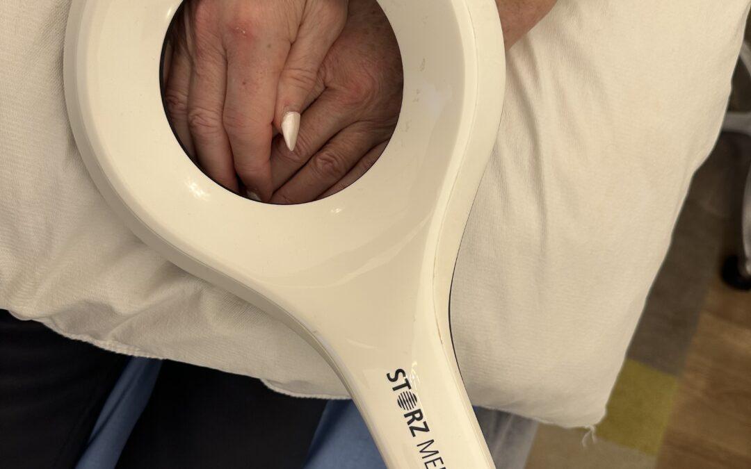
Painful Thumb: Here is a brilliant thumb brace for all kinds of aches of the thumb

January 26th, 2012

Excellent protection to pain and discomfort of your over worked thumb


A brand new CMC thumb brace designed and developed by eminent hand therapists Judy Colditz and Nettie Koekebakker.

Pain of the thumb and osteoarthritis is a common thumb disorder. It is a type of ‘wear and tear’ of the articular cartilage, affecting in particular people between 40 and 60 years of age.The Push CMC is a slim and compact product. The brace can be used in the water. Hygiene is important around the hand. Therefore the synthetic material is antibacterial and the brace can be washed in a washing machine at 40°.


This simple clever thumb brace is outstanding,  The thumb joint over the years  is prone to disabling pain which prevents one opening normal screw top bottles, jars tins and all kinds of other essential normal housemaking and job activities.  What most people do not know is that if the ligaments of the thumb are not protected from many of our normal daily activities the fine muscles of our thumb waste away and atrophy leaving to further weakness and permanent dysfunction.

By where this small neat thumb brace, you can get on with your daily activity, keeping your muscles active and therefore not wasting away while the inflammation and ligaments settle down.  For those of you with mild OA of the thumb you can wear the thumb as a temporary protection when you want to do some more heavy activity such as gardening or house hold work.

I had had excellent positive feedback from those who have purchased this neat thumb brace and I have used it for my own thumb which is now  completely pain free.   Taking some types of pain killers actually mean that you damage your joints and ligaments even more or some NSAIDS actually delay or stop the healing of collagen tissue.

Here is the link where you can buy this thumb brace for yourselves, I have added the link to facebook as well  where I add excellent you tube videos and other good sources of better management of all kinds of aches and pains.  Please do go to my facebook link as Facebook is so clever and it looks much neater then my blog.


Here is the reviews of this clinic starting from 2006 –    Reviews are increasingly important in this economic climate .


Helen How

Registered Osteopath

14 Craighall Gardens

Edinburgh EH6 4RJ


Back Pain and Core Stability – Stuart McGill shows how to train your Core Muscles with further damaging your lumbar joint facets or increasing sciatic pain

January 13th, 2012

One of the main reasons for the reviews on this clinic are genuine, realistic and charming is thanks to one of my favourite Medical Research is thanks to the years of research by Stuart McGill

I am delighted to see that now Stuart McGill has now some excellent videos on how you can seriously damage your back with excessive exercises on gym equipment (if you wish) or how to exercise safely.


As Stuart says most people will suffer from some back pain at time in their lives

Please look and understand this excellent video    One has to keep the spine stiff rather than increase the range of movement as this causes damage on the muscles ligaments and lumbar vertebrae causes chronic inflammation and long term pain.  The wrong movements abuse the back and increase your chronic pain.  Prolonged sitting makes your back muscles weak and much more prone to back injury which is why long term back pain in on the increase

Here is another of Stuart McGill`s videos discussing on how to train you core properly.

Helen How

Registered Osteopath

14 Craighall Gardens

Edinburgh EH6 4RJ

Fees £35.00 for hour  and for simple follow up cases £20 for half an hour.

I have kept to these comfortable affordable fees for years as I find it takes one hour for many of the more challenging complicated cases.


Arsenal Football Club SEMS Conference 2012

December 28th, 2011

As a practising Osteopath who qualified in 1981 and over the years one had attended many Sports Medicine Conferences  I would never have dreamed that one day I would be able to visit and be given a tour of the Emirates Stadium thanks to the open hospitality of Arsène Wenger and the Emirates for their world class Rehabilitation Centre.


Arsenal Football Club, Emirates Stadium, London
Meeting Programme
0800 Registration, Coffee and Commercial Exhibition
0815 Tour of Emirates Stadium
0930 Welcome and Opening
Mr Arsène Wenger, Arsenal FC Manager
Dr Gary O’Driscoll, Arsenal FC Medical Director
Morning Session Chair Dr Gary O’Driscoll
0940 New treatment methods for the chronic painful Achilles and patellar tendon allowing for a fast return to sports
Professor Hakan Alfredson
1020 Supplements, food and positive doping tests
Professor Ron Maughan
1100 The Talent Myth and its implications
Mr Matthew Syed
1140 Refreshments and Commercial Exhibition
1140 The current use of prolotherapy in acute injuries
Adam Mitchell
1220 The Athlete’s Hip – An Arthroscopist’s View
Richard Villar
1300 Question Session
All speakers
1330 Lunch and Commercial Exhibition
1340 Tour of Emirates Stadium (30 minutes – optional)
Arsenal Football Club SEMS Conference 2012
125 years in elite sports medicine; what challenges lie ahead?


Helen How

Registered Osteopath

14 Craighall Gardens

Edinburgh EH6 4RJ

0131 551 1044

Fees £35.00 for one hour with easy parking.

Here are reviews and feedback  about this Clinic different reviews are on Google a nd others in Edinburghonline.    Feedback has become part of our Professional Revalidation Guidelines for Osteopathy.

www.howclinic.com


Back Pain

November 10th, 2011

As a Registered  Osteopath looking after back pain  for over 25 years with advice, help, and patient feedback and attending many Medical and healthcare courses Where the Doctors, physiotherapists, chiropractors nurse and many others are  all attempting to   find better ways of managing and reducing chronic back pain.  Back pain is complicated as muscles, tendons, ligaments and bones get a bit worn out and inflamed.  One has to trigger the healing and repairing of these inflammations using huge variety of management plans and lots and lots of feedback from you the patient.  That is why there is an enormous number of scientists, researchers, clinicians and statics involved is collecting the knowledge, evidence, skills and communicating it effectively to the patients throughout the NHS,

I think we all forget how far the diagnosis of back pain has progressed with the most up to date MRI scanners being able to image in much great detail depending on the make of the scanner but ask your Radiologist.   Imaging is often the first investigation that your GP or Consultant will arrange if you have severe, chronic and disabling back pain.  As the end of maybe the investigations, sometimes surgery, pain relief there is the ongoing management of your own personal back.

One the facebook section of this website you will see some brilliant clinical and educational websites from various parts of the world including Great Britain


Please do read some of this clinics  reviews  which were started in 2006 by patient.  Over the years with the scientific evidence based publications by Mark Comerford, Professor Stuart McGill, Professor Tim Watson and many other acknowledgements such as the British Sports and Exercise Medicine and Professor Nicola Maffulli to help everyone who has attended this Clinic.  Thank them for all the hours of research and world wide lecturing.


I use Face book on the first page of my website for educational purposes so there are the excellent YOU Tube videos on lumbar disc pressure, Professor Stuart McGill shows his research and safe Core Exercises which is why these reviews are so kind on my Clinic.    Here is one of Stuart McGill`s videos.    Another essential video to watch if you want to really understand how maintain a happy long term healthy back. Click Here.   Even patient who have had chronic pain for over 7 years have greatly improved by following Stuart McGill`s brilliant research.   There are many strange myths and misleading information and many exercises are more harmful then beneficial so take care and be sensible

Here is my last review from Google.  I put the reviews here more because I recently bought a cash register from a London based website and it was a terrible experience because they sent the wrong cash register and when it finally arrived I could not operate it because cash registers are very complicated systems.  I was meant to spend another £200 for the company to send up representative to teach me how to use this cash register.  I still cannot use it properly but I can give out your receipts now.  In the end I had to call another company who had an excellent customer care line and we looked at their you tube video instructions.


Wendy ‎ - 21 Nov 2011

Genuinely caring, professional and excellent value. My husband and I visitedEdinburgh on our anniversary and I arrived in pain! After searching for a local physiotherapist / chiropractor /osteopath on my smartphone, I read the wonderful Google reviews and decided to give OsteopathHelenHow at How Clinic a call. Never been to an Osteopath before, but Helen was very different to other practitioners I had seen. She took the time to understand and treat my back pain as well as educate me how to prevent it recurring. She used a massage machine and ultrasound as well as a couple of different treatment methods, and I really felt the difference. As well as an expert in her field, Helen’s also a really lovely lady and her treatment is also excellent value for money compared to others I have seen. Would loved to have continued treatment but unfortunately we don’t live in Scotland! Finding someone with the same caring and professional approach where I live is not going to be easy!!


Below I have included some excellent videos to help you understand the complexities of back pain


Here is some of the excellent video links which are useful if you are researching more about your back pain.


The Costs :-       Over the year the average number of sessions and now that I am on the revalidation pilot scheme, all Osteopaths have to collect this evidence is between 1 to 4 sessions.   The number of sessions has greatly reduced at my Clinic with the introduction of the new effective levels of Ultra Sound treatment which takes between 5 to 20 minutes to apply

There for if you attend for 4 sessions in the year that comes to a total cost of this Clinic of £140.00

Some of my clients have attended therapists for years without the application of Ultra Sound and the feedback is that the Ultra Sound is helping them to resolve the body`s ability to repair and heal.  However it is important that give time for the body to heal so hence one has to be sensible.

I hope that helps you understand why some of the reviews for this Clinic are good


Helen How
Registered Osteopath
14 Craighall Gardens
Edinburgh
EH6 4 RJ
0131 551 1044
£35 all included per hour
Easy parking

Sports Medicine & The Elite Golfer Sponsored by the PGA.

November 5th, 2011

I was lucky to able  attend the  first ever Sports Medicine Course and the Elite Golfer sponsored by  PGA European Tour. which was held at the Rudding Park Hotel Harrogate on Thursday 3rd November.  It was the idea  of  Dr P J O`Connor who is an experienced sports radiologist and is imaging lead for the 2012 Olympics and advisor to the BGA European Tour.  I have listed below the educational websites which give you an insight into the most current up date medical evidence based management of the common conditions such a tennis elbow, low back pain and wrist pain which is seen in both the professional and the amateur golfer.  I strongely recommend you take some time to read the websites as they are outstanding in their knowledge and expertise.

The Medical team kindly opened it as multi displinary which allowed physiotherapists, chiropractors, doctors and other sports care professional to attend.

The dynamics of the golf swing has changed and evolved radically over the past 10 years and before the more natural slower golf swing would appear to be much safer and less stressful to the  wrist,  shoulder,hand and back.

With the speed of the golf swing over 130 mph in the profession golf player the number of repetitive injuries has significally increased and therefore it has become essential for the professional player to have ready access to high quality imaging and consultant radiologists and specialists in the back, elbow, shoulders, hands and wrist.

Here are some of the websites of the medical specialist for your own educational purposes.

  1. Research and MRI Scans and all the other forms of imaging everything you need to know and understand about the elbow can be found on ElbowDoc.co.cuk . Elbow Pain
  2. Golf Fitness and strengthening exercises by the PGA to protect your low back 
  3. Diagnose your own shoulder pain on ShoulderDoc.co.uk 
  4. Do you understand the X – Factor in your golf swing  – here is an insight from the PGA Website

Helen How

Registered Osteopath

0131 551 1044

£35 per hour  all inclusive

Read reviews

Reviews on Osteopathy and Osteopaths

October 22nd, 2011

Over the past year the feedback from patients in Edinburgh  is that they want to read more current real reviews  from the patients attending their  Osteopath, Chiropractor and Physiotherapist . This is because Amazon and Trip Advisor have been so successful in customer care reviews that we all now have expect to read reviews about all kinds of subjects.

I, myself, bought my cash registered recently on the internet and did my research except the particular website did not have any customer reviews and my cash registered was sent late, with the wrong parts ordered and then I discovered that the instruction manual is exceptionally difficult to understand.   This Cash Registered Company was going to charge me £200 to send up someone to explain to me who to use my cash register.  I still do not really understand how to use it properly but I collect the invoices and receipts as required by our Tax Regulations.


Ultra Sound Machine working on a patient

This week two patients both expressed a wish that there was a single website which supported reviews on all of us “Therapists” provided the reviews were seriously genuine.    It is true that there is a wide range of approaches, treatments and costs of fees and thoughts on mangement principles available all through the Great Britain.  As I am participating in the Pilot Revalidation Scheme , as Osteopaths we now have to have within our Practice Management, feedback forms, complaint forms, and peer review supervision.  I am spending extra hours reviewing and changing my own management practice and it  is time consuming.

One of the biggest surprises to me it the wish to see more current reviews for patients attending a particular clinic or Osteopath  rather then a   promotional based  website.

Surprisingly, I use twitter and facebook on my website more because I have found that both twitter and facebook can be used to insert excellent educational videos and links for speedy efficient open access rather then the more duller reading of a blog.  The blog seems to be reassuring that the Osteopath or who ever is participating and reading up more scientific or educational material to further the Osteopath`s own knowledge and professional skills.


My first  review started was started by C. Samson  in 2006 and patients have kindly provided 15 more on my current interests and practice management  please read their reviews on Edinburghonline

How Clinic 

Helen How

Registered Osteopath

14 Craighall Gardens

Edinburgh EH6 4RJ

Contact via email

0131 551 1044


Osteopaths on the Pilot Revalidation

October 19th, 2011

As an Edinburgh based  Osteopath participating in the Pilot Revalidation scheme  which is being supervised by our General Osteopathic Council and KPMG. we  have to collect evidence, documentation and permission from a few selected patients and also ask fellow Osteopaths to come in and peer review and give us feedback.

Helen has been a Registered Osteopath, Trained at the European School of Osteopathy and graduated in 1981.   As patients are  feedbacking  requests  to read views about  Osteopaths  here are Helen`s  reviews on Edinburgh on line

The Osteopaths have been given four revalidation Themes

  1. Communication and patient partnership
  2. Knowledge skills and performance
  3. Safety and quality in practice
  4. Professionalism

The General Osteopathic Council has given an overview of development of the revalidation scheme and the next steps in development.
Department of Health has the following expectations as described in the following paragraph.

In November 2010 the Department of Health wrote to the chief executives of all of the non-medical healthcare regulators setting out their current expectations in relation to the development of revalidation. Health ministers in the four countries of the UK have agreed that the healthcare professions regulators should “continue to gather the evidence base to assess the feasibility and proportionality of non-medical revalidation.” This means that we will be continuing with the developmental work that we are doing on revalidation. Click here to read the letter in full.

At the same time, the Department of Health released additional funding to the GOsC to support our revalidation pilot and to ensure that the proposed scheme is both proportionate and supported by robust analysis of the costs and benefits. This is good news for osteopaths as it will allow us to make effective progress in developing the scheme, while the grant of £167,000 means to develop our pilot scheme


Feedback from the pilot will be collated and analysed and will feed into a further consultation on revalidation. Depending on the results of the independent analyses and the consultation results, we plan to implement revalidation in 2014. However, the nature of this large project means that we must be very thorough, to ensure that the scheme meets the needs of osteopaths and patients. This means that timescales are flexible.

Helen How

How Clinic

Registered Osteopath

14 Craighall Gardens

Edinburgh EH6 4RJ

0131 551 1044

Fees £35.00 for one hour including consultation.

Simple follow up £20.00 for 30 mins

Pregnancy – the Aches and Pains

October 16th, 2011

As an Osteopath who has had 3 children and has worked with Pregnancy, babies and children now for over 30 years. The most important news is that on 3rd October 2011 the state-of-the-art Birth Centre at the New Royal Edinburgh Infirmary has six delivery roooms all with en-suite facilities. Each Room is bright, spacious modern designed rooms with a birthing pool and equipment to enable you to have the most comfortable birth experience complete with mats, pillows and birthing balls. I have heard wonderful feedback already which is most welcoming

Brief Background on Helen

Helen has been a Registered Osteopath, Trained at the European School of Osteopathy and graduated in 1981.   Helen is participating in the Pilot Revalidation Scheme  in which  patients   requested   to read  current reviews about  Osteopaths  in Edinburgh.  Here is the link to  Helen`s  reviews on Edinburgh on line


I have found this excellent article written by Core Concepts a team of physiotherapists based in Singapore on the Aches and Pains of Pregnancy. I had added a few easy tips.

Pregnancy – the Aches and Pains by Core Concepts

If you are pregnant and feel your back or pelvic pains more often than usual, you are not alone. Studies have shown that more than two-thirds of pregnant women experience back pain and almost one-fifth experience pelvic pain2,.3. As pregnancy progresses, the intensity of pain may increase and interfere with your work, daily activities and sleep.

Causes my back and pelvic pains

As your pregnancy advances, your back experiences more strain from three key changes – a shift in your centre of gravity, hormonal changes and abdominal muscles changes.

Centre of Gravity Shift

Your centre of gravity shifts forward due to the growing bump. This accentuates the curvature of the spine and adds stress to the spinal joints, contributing to the tightening of your spinal muscles. The growing bump also adds extra load on joints, and that means more work for the back muscles, which is why your back pain may worsen as the day progresses as they tire.

Hormonal Changes

Relaxin, a hormone produced during pregnancy, acts on ligaments making them suppler. The ligaments that hold the pelvis bones together gradually loosen to prepare you for labour and birth. Unfortunately, this compromises joint stability of the pelvis. Making you feel less steady and cause pain when you sit for long periods, stand, walk, roll over in bed, get out of a low chair, bend, and lift.

Abdominal Muscles Changes

The expanding size of the uterus stretches the abdominal muscles reducing support for the spine. Diastasis recti, a pregnancy-induced condition, is the separation of the rectus abdominis muscles in the middle, reducing the ability of the abdominal muscles to support the pelvis and spine1.

Common areas of back pain during pregnancy

Expecting mother most typically experience low back (lumbar pain) and posterior pelvic or sacroiliac back pain.

With low back pain, you feel it across the lower spine, near or at the level of your waist. Prolonged sitting or standing usually makes it worse, so does wearing high heels. It tends to be more intense towards the end of the day.

Posterior pelvic pain is typically felt at or near the 2 dimples at the back of your pelvis (hip). You feel the pain deep inside the buttocks or in the back of your thighs on one or both sides. Activities that aggravate the pain include walking, climbing stairs, resting on one leg, getting in and out of a low chair, rolling over and twisting in bed, and lifting.

What can I do to prevent back pain?

If you have a history of back pain, you are more likely to get it again. You are also prone to back pains if you lead a sedentary lifestyle and have weak back and abdominal muscles with poor Core Stability

One of my best tips is very simple, if you are really struggling use a small walking stick to help you get up and down out of your chair or if you are getting really pubic stretching pain – see if you can borrow a pair crutches to help you around the home. It does not matter if any one sees you at this stage because you will have limiting the stretching and stressing of the ligaments and low back muscles


To prevent recurrence, start on an exercise program to stretch and strengthen muscles that support the back, which include your abdominals. Remember first to get clearance from your doctor before you start any exercise program.

Adopt good postures in your daily life will do wonders for your back.
For example,

• Standing. Stand up straight. This gets harder to do as pregnancy progresses, but try to keep your chin tucked in, drawing your shoulders back and down with the tummy and bottom tucked in. Expectant women tend to slump their shoulders and arch their backs as their bumps grow, which puts more strain on the spine.

• Sitting. Try to avoid prolong sitting and get up take frequent breaks. Support your feet with a footstool can help reduce back pain, and using small pillow to support your lower back helps too.

• Lifting. Bend at your knees, keeping your back straight and lift using the strong thigh muscles instead of the small muscles of the back. Do not twist as you are lifting. Get help for heavy objects.

• Sleeping. To rest better at night, sleep on your side with one or both knees bent and a pillow between your legs. As your pregnancy advances, use a rolled up towel or small pillow to support your tummy.

Finally yet importantly, pay attention to your body. If you find that a particular activity or exercise aggravates your pain, stop doing it. Ask your osteopath or physiotherapist whenever in doubt!

Helen How
Registered Osteopath
14 Craighall Gardens
Edinburgh EH6 4RJ

0131 551 1044

£35 per hour


Knee Pain

October 12th, 2011

Frontal Knee Pain is one of the most commonest conditions I see as an Osteopath here in Edinburgh. This is the pain you feel when you walk up and down stairs and you think it is going to improve but niggles and niggles on. The frontal pain comes from your knee cap constantly moving up and down as you bend your knee and your front muscles of your thigh (the quadriceps) become over strong and tight contract the knee cap into tracking groove of your knee.

Helen How  Registered Osteopath, 14 Craighall Gardens, Edinburgh EH6 4RJ.  See reviews her on Edinburgh on line


Treatment of this prolonged painful condition must include increasing the long term flexibility of the front and back muscles of your legs so that the knee cap or patella is not constantly irritating the boney cartilageous surface of the knee. These muscles can become very tight and need stronger techniques which such as my fleximatic machine to really release those tight muscles and then one needs to stimulate the inflammation to settle with a good effective dose of Ultra Sound. Please read further the good article which includes the other combination of treatments which may be needed to resolve frontal knee pain.


This below article comes from Wilkepidia and gives you a good back ground education and advice on positive treatment.

Patellofemoral pain syndrome (PFPS) is a syndrome characterized by pain or discomfort seemingly originating from the contact of the posterior surface of the patella (back of the kneecap) with the femur (thigh bone). It is the most frequently encountered diagnosis in sports medicine clinics.
Contents

1 Mechanism
2 Treatment
2.1 Exercises
2.2 Rest
2.3 Ice and medication
2.4 Taping and braces
2.5 Arch support
Mechanism

The cause of pain and dysfunction often results from either abnormal forces (e.g. increased pull of the lateral quadricep retinaculum with acute or chronic lateral PF subluxation/dislocation) or prolonged repetitive compressive or shearing forces (running or jumping) on the PF joint. The result is thinning and softening (chondromalacia) of the articular cartilage under the patella and/or on the medial or lateral femoral condyles, synovial irritation and inflammation and subchondral bony changes in the distal femur or patella known as “bone bruises”. Secondary causes of PF Syndrome are fractures, internal knee derangement, OA of the knee and bony tumors in or around the knee.

Specific populations at high risk of primary Patellofemoral Syndrome include runners, basketball players, young athletes and females especially those who have an increased angle of genu valgus (aka “Q-Angle” or commonly referred to as “knock-knees”). Typically patients will complain of localized anterior knee pain which is exacerbated by sports, walking, sitting for a long time, or stair climbing. Descending stairs may be worse than ascending. Unless there is an underlying pathology in the knee, swelling is usually mild to nil. Palpation, as well, is usually unremarkable.
Treatment
Exercises

Quadriceps strengthening is commonly suggested because the quadricep muscles help to stabilize the patella. Proper form is very important. Inflexibility has often been cited as a source of patellofemoral pain syndrome. Stretching of the hip, hamstring, calf, and iliotibial band may help restore proper biomechanics Furthermore, the use of a foam roller may help to add flexibility and relieve pain from sore or stiff muscles in the leg.
Rest

Patellofemoral pain syndrome may also result from overuse or overload of the PF joint. For this reason, knee activity should be reduced until the pain is resolved. Those with pain originating from sitting too long should straighten the leg or walk periodically. Those who engage in high impact activity such as running should consider a nonimpact activity such as swimming or aerobics on an elliptical machine.
[edit] Ice and medication

To reduce inflammation, ice can be applied to the PF joint after an activity. The ice should be kept in place for 5 minutes only but can be applied at least 5 to 6 times in a day
Taping and braces

In addition to physical therapy, external devices such as braces and tape could be used to stabilize the knee. These devices will not correct the underlying source but may prevent further injury. For this reason, they should be used in conjunction with and not in lieu of physical therapy. The technique of McConnell taping has been helpful in some studies
Arch support


Low arches can cause overpronation or the feet to roll inward too much increasing the Q angle and genu valgus. Poor lower extremity biomechanics may cause stress on the knees and ultimately patellofemoral pain syndrome. Stability or motion control shoes are designed for people with pronation issues. Arch supports and custom orthotics may also help to improve lower extremity biomechanics.

Ultra Sound is beneficial to stimulate the recovery and healing of the ligaments and other soft tissues

Helen How
Registered Osteopath
14 Craighall Gardens
Edinburgh EH6 4RJ

0131 551 1044

As the Femoral Patella Pain requires 20 minutes of loosening up off your quadriceps and hamstrings then Ultra Sound and checking your foot ware and orthortics I find I need the full hour for this condition and it my normal rate of £35 per hour.

See reviews on Helen in Edinburghonline


Shoulder Pain

October 10th, 2011

Helen How  Registered Osteopath, 14 Craighall Gardens, Edinburgh EH6 4RJ.  See reviews her on Edinburgh on line

This is a surprisingly common cause of Shoulder Pain which is surprisingly difficult to treat and if you glance through this lengthy article to be found in Patient.Uk.Co I have been working on better management solutions with good feedback and participation from the clients. Osteopathy has worked with different shoulder pains but the biceps tendon is a thick tendon which takes a great deal of mechanical loading during the whole day.  It is therefore really rather difficult to treat and most of the suggested treatments seem to be very slow or not really work unless you sit around resting and relaxing all day.

Here from the Sports and Exercise Medicine Course Sponsored by the PGA  in Harrogate a brilliant Article on Diagnose your Shoulder   ShoulderDoc.com

If you continue to be interested then here the Medical Rehabilitation Stepped Intensity Exercises which have are comprehensive however please do not attempt any of these exercises without asking your surgeon or rehabilitation specialist as diagnosis and patient compliance is essential.  Shoulders are highly complicated structions and can easily be made worse.  ShoulderDoc.co.UK Shoulder Rehabilitation Exercises


I have now managed over 6 cases of biceps tendonopathy this year and have found that it responds well to  electrotherapy and protective  strapping of the tendon and shoulder.  The shoulder strapping takes the loading off the biceps tendon and with promotion of the sound waves from the ultra sound the cells which clear up inflammation and heal the damaged tendons have a window box opportunity to get on with their healing.   So far all cases have responded and they can all sleep soundly through the night without being disturbed by pain.

Biceps Tendonitis

Biceps tendonitis occurs in the long head of the biceps tendon as it runs on the anterior aspect of the humerus between the attachments of the supraspinatus (greater tuberosity) and subscapularis (lesser tuberosity). The function of the biceps brachii muscle is supination and flexion of the forearm. Problems with the biceps tendon can result from impingement or from inflammation. Biceps tendonitis can also occur secondary to compensation for other shoulder disorders, particularly rotator cuff disorders, labral tears, and intra-articular pathology.1

The diagnosis can be difficult not just because there are many other causes of shoulder pain, but also because it is common for several different problems to exist in the same shoulder and contribute to a sometimes confusing clinical picture.
Injuries appear to occur more often among patients who engage in frequent pulling, lifting, reaching or throwing (work or recreation). It occurs typically with repetitive overhead activity.

Complications are more common in older patients, particularly damage and rupture of the tendon.
Clinical series have described biceps tendon ruptures in rock climbers and weight lifters.
Pathophysiology

Historically, all disorders of the biceps tendon were described as biceps tendonitis. However, degenerative changes occur in the tendon without inflammation. An inflammatory pathology may explain the pain experienced in the biceps tendon. The different terms used to describe the pathophysiology more accurately are defined below:2

Tendonitis describes inflammation of the tendon and the paratendon. Chronic overload is thought to cause microscopic tears in the tendon which trigger an inflammatory response.
Peritendinitis describes inflammation of the paratendon or tendon sheath. This usually results from a direct injury or irritation caused by impingement of the tendon against a bony prominence. This is also described as a tenosynovitis.
Tendinosis is an histological definition and describes degenerative changes in the tendon:
Macroscopically there is a degenerative tendon with disorganised tissue (mucoid degeneration).
Microscopically there are degenerative changes to the collagen with fibrosis. Inflammatory mediators are not usually present in tendinosis. Older injuries (more than 3 months) have less inflammation and more degenerative change.
The term tendinopathy refers to the clinical presentation of a symptomatic tendon rather than the underlying pathology (degenerative or inflammatory). Three aetiological theories for tendinopathy are described:
A mechanical theory where repetitive loading of the tendon causes microscopic degeneration. Fibroplasia occurs within the tendon, resulting in scar tissue.
A vascular theory describing tendon degeneration with secondary areas of focal vascular disruption.
Neural modulation. This is a new theory proposing that tendinopathy arises from neurally mediated mast cell degranulation and release of substance P.

Epidemiology

The incidence of biceps tendon injury in sport and different occupations is unknown.3
Biceps tendonitis occurs in a variety of sports including weight lifting, tennis, wheelchair athletics (and general wheelchair use), cricket, baseball, kayaking and other sports where overhead activity is involved.4,5,6
Degenerative tendinosis and biceps tendon rupture are usually seen in older patients.
Isolated tendonitis often presents in young or middle-aged patients but the exact incidence is unknown.

Presentation
History

It is important to consider some basic points in the history, as these have an important bearing on the management, including use of injections and whether or not prompt referral is appropriate.

General factors:
How old is the patient? Older patients are more prone to impingement, tendinosis and tendon rupture.
Are there significant comorbidities? Rheumatoid arthritis is more often associated with tendon rupture and significant joint pathology.
What is the patient’s occupation?
What are their hobbies?
What is the site and nature of the pain?
Typically, it is a vague anterior shoulder pain which has an aching quality.
It is aggravated by lifting (and especially pulling and pushing overhead) and relieved by rest.
Is the pain acute or chronic? If the pain has been going on for longer than 2 months it is likely that there are degenerative changes in the tendon. Referral is more appropriate. Tendon rupture is more likely.
Has there been a tendon rupture? What was the mechanism of any injury involved?
Usually there is no history of injury but, if there is such a history, beware of possible tendon rupture.
With rupture of the long head of the biceps tendon patients may report a sudden and painful popping sensation with appearance of the “Popeye” deformity in the anterior upper arm (bulging retracted muscle).
When there has been a traumatic injury, the biceps tendon rupture is often preceded by a history of shoulder pain that resolves following a painful and audible snap.
Has overuse been a factor?
Bicipital tendonitis frequently occurs from overuse syndromes of the shoulder.
Biceps tendonitis tends to occur with repetitive overhead activity which may be sporting, recreational or occupational.
These are common in athletes where, again, repetitive overhead activity is involved. Examples of such sports include cricket, swimming, racquet sports, weight lifting, rowing and kayaking.
Are there other associated symptoms?
Shoulder instability and subluxation can be associated with biceps’ degeneration associated with chronic tendonitis.
Instability and subluxation also occur with labral tears, often with additional locking or catching symptoms.

Examination

Diagnosis requires a thorough shoulder examination. Essentially the assessment should locate the site of tenderness and demonstrate which movements aggravate the pain.

First know and consider function and anatomical landmarks:
The long tendon of the biceps runs along the anterior aspect of the humerus between the attachments of the supraspinatus (greater tuberosity) and subscapularis (lesser tuberosity).
Between the lesser and greater tuberosities is a ligament which retains the long biceps tendon which runs in a groove.
The long head of biceps inserts at the upper edge of the glenoid labrum and supraglenoid tubercle. The long head of the biceps tendon helps to stabilise the humeral head, especially during abduction and external rotation.
The biceps is a powerful supinator and flexor of the forearm.
The tendon may dislocate from this groove or become inflamed.
Inspection:
The muscle bulk of the shoulder girdle, looking for wasting.
For any anatomical abnormalities.
For postural deformity.
Palpation:
Local tenderness is usually present over the bicipital groove (typically located 3 inches below the anterior acromion).
Compare one side with the other because there is often some tenderness on the unaffected side. The affected side will be more tender.
Examine for point of tenderness with the arm in 10° of external rotation. The special tests for biceps tendonitis below essentially elicit tenderness at this site by various means.
Examine the range of movement:
Bending the elbow and abduction of the shoulder may cause pain up to the shoulder.
Perform a neurological examination:
Briefly check power, tone and reflexes. There will usually be no neurological deficit.
Muscle power may be limited by pain.
Use special tests to confirm the diagnosis. A number have been described. Detailed description is beyond the scope of this article but it is worth being aware of their significance as they may be described in reports and letters. These are:
Biceps tests, including Speed’s tests7 (1 and 2 to confirm biceps tendonitis), Yergason’s test (not considered universally useful)7, Gilchrist’s test (use of weights to confirm biceps tendonitis) and the Lippman test (test for tenosynovitis with instability of the biceps tendon).
Other tests looking for rotator cuff, labral and acromioclavicular joint pathology. These include the Hawkins-Kennedy test, and the Neer test (acromioclavicular joint) and O’Brien’s test, anterior slide and clunk test (labral tests).

Investigation

This is a clinical diagnosis and investigation is not routinely required. However, investigations may occasionally be useful – for example, when pain is severe, the diagnosis is in doubt or functional limitation is marked.

Ultrasound is the examination of choice.8,9 Soft tissue ultrasound may not be easily available in primary care but can help in the diagnosis and exclude degenerative disease of the tendon prior to injection.
Plain X-ray may be used when there is a suspicion of neoplasia. The demonstration of spurs, calcification or changes of osteoarthritis is unlikely to help management.
MRI scan can demonstrate the whole course of the biceps tendon (including the intra-articular tendon and related intra-articular pathology).10,11 However, it is not appropriate or cost-effective for routine use. It is indicated after unsuccessful rehabilitation or where there is suspected rotator cuff or labral tear injury.
Injection of local anaesthetic may be used to confirm the diagnosis, and not just for therapeutic reasons.12 Relief of symptoms helps differentiate biceps tendonitis from, for example, referred rotator cuff pain and glenohumeral joint disease.

Differential diagnosis

Injury:
Glenoid labrum tear (anterior)
Fractures (greater or lesser tuberosity)
Glenohumeral instability (humeral subluxation)
Subscapularis strain or tear
Inflammation:
Synovitis
Capsulitis (frozen shoulder)
Inflammatory arthropathy
Others:
Neoplasm
Peripheral nerve entrapment

Management1

The appropriate management will depend on the patient and the length of history.
Treatment should incorporate:

Rest from lifting, stretching and overhead use of the affected arm.
Ice applied for 10 to 15 minutes three to four times per day for the first 2 days.
Non-steroidal anti-inflammatory drugs taken regularly for pain over the first few weeks.
Other modalities of treatment may be employed by physiotherapists, including ultrasound, transcutaneous electrical nerve stimulation and gentle stretching exercises.
Local anaesthetic and steroid injection is typically recommended 3-6 weeks after the acute injury (see below).
Orthopaedic referral should be considered if after 2 months the patient’s symptoms persist. If biceps tendon rupture is suspected then early referral is appropriate, especially in patients under the age of 40 years.

Injection

Many people use methylprednisolone or triamcinolone that are available already mixed with local anaesthetic. Although this is convenient, these strong steroids can cause fat atrophy and depigmentation in the skin over the injection site and so it is often argued that hydrocortisone with lidocaine should be used in preference and the stronger steroids saved for deeper injections.13With the patient sitting or lying, the biceps tendon is identified in the groove, and the point of insertion noted. To inject into the area of the long head of the biceps tendon, the needle is inserted directly into the most tender area over the bicipital groove. The needle should enter the skin at 30° and be directed parallel to the groove. The objective is to infiltrate the area in and around the groove and not into the tendon, as this may result in rupture. Increased resistance to the injection suggests that the needle is in the tendon and it should be withdrawn a little.

The risk of tendon rupture is increased with repeat injections and also in patients over 40 years of age.

As with the other injections, shortly after performing it, repeat physical examination, and pain and restriction of movement should have disappeared.
Advice after injection

The patient may be impressed by the instant cure but warn that:

The initial benefit is from the local anaesthetic and its presence assures that the injection was put in the right place. However, it will wear off over the next 2 or 3 hours.
The benefit of the injection takes 2 or 3 days.
Sometimes the pain becomes worse than it was before it gets better.
Rarely, crystals of steroid precipitate and cause extreme pain. This may still precede cure.
The patient should not forget what caused the lesion initially and should return slowly to full activity. Restrict lifting and overhead activities by the patient for 30 days after the injection.

Surgery

Surgical intervention is rarely indicated. It may be appropriate for partial rupture of tendons and is usually performed early (under 6 weeks).
If there is slow and gradual improvement, surgical intervention is not recommended for bicipital tendonitis but may occasionally be indicated if a trial of conservative care for 6 months is unsuccessful. Acromioplasty with anterior acromionectomy is the standard surgical treatment for bicipital tendonitis, although arthroscopic decompression is also performed.
Prevention

Biceps tendonitis tends to occur with repetitive overhead activity. Modification of activities may be appropriate with certain occupations with guidance from a physiotherapist. In addition to physiotherapy, advice from a sports physician may be sought where bicipital tendonitis may relate to technique which may be modified with suitable coaching advice.
Document references


Helen How

Registered Osteopath

`14 Craighall Gardens

Edinburgh EH6 4RJ

£35 per hour and/or £20 for simple follow up sessions


0131 551 1044

see reviews on Helen on Edinburghonline